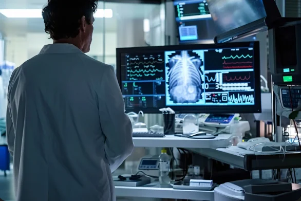
Medical Imaging
Overview
Job Growth
N/A
Duration
N/A
Avg. Salary
N/A
Career Paths
N/A
Program Description
Medical Imaging trains students to use medical imaging equipment to help diagnose and treat patients. You will learn anatomy, radiographic physics, imaging techniques (X-ray, ultrasound, CT basics), radiation safety, and patient care. The program combines classroom theory with hands-on practice in imaging labs and hospital placements. Graduates can work as radiographers, sonographers, imaging technicians, or assistants in hospitals, clinics, and diagnostic centres — with opportunities to specialise later in CT, MRI or ultrasound. This program is practical, rewarding, and in demand across Ghana’s healthcare system. If you enjoy science, patient care and technology, Medical Imaging opens a clear path to a respected health career with real impact on patient care and community health.
Aims & Objectives
Develop safe patient-centred imaging skills to perform standard X-ray and basic ultrasound procedures under supervision.
Master radiation protection and infection control techniques to maintain ALARA standards in clinical settings.
Understand human anatomy and radiographic anatomy to produce diagnostically useful images.
Perform image quality assessments and apply basic digital image processing and PACS workflows accurately.
Record and manage patient data and imaging reports using proper medical documentation and health information systems.
Why Choose This Program?
Strong Job Demand
Ghana’s growing healthcare services and diagnostic centres need trained imaging staff, offering steady employment and career stability.
Hands-on Training
Practical lab sessions and clinical placements give real experience operating imaging equipment and caring for patients.
Clear Career Pathways
Start as a radiographer or sonographer and specialise later in CT, MRI, or ultrasound with further training and certification.
Transferable Technical Skills
Skills in imaging equipment, PACS, and patient assessment are useful across hospitals, private clinics, and health programmes.
Industry Connections & Internships
Training programmes often include placements in major teaching hospitals and diagnostic centres, helping build professional networks.
Skills & Tools
Skills You'll Develop
Evaluate patient needs and correctly position patients for X-ray and ultrasound exams to ensure diagnostic image quality.
Operate digital X-ray units, ultrasound machines, and image capture systems according to manufacturer and safety protocols.
Apply ALARA principles, use protective equipment, and follow sterilisation procedures to protect patients and staff.
Manage images and reports using PACS/DICOM viewers and enter accurate patient data into electronic health records.
Assess and optimise image contrast, exposure, and positioning; perform basic post-processing to enhance diagnostic value.
Tools & Resources
PACS (Picture Archiving and Communication System)
DICOM viewers (radiology image viewers)
Electronic Health Records (EHR) systems
Radiology Information System (RIS)
Challenges & Tips
Challenges
Complex science and physics concepts (radiation physics, anatomy).
Balancing theory with practical skill learning.
Tips & Advice
Break topics into short study sessions, use visual resources (anatomy charts, Radiopaedia) and join study groups.
Practice regularly in labs, record procedures, ask supervisors for extra supervised practice during placements.
Video Guide
Frequently Asked Questions
Ready to Apply?
Find programs that match your grades and interests - even if you haven't written WASSCE yet
Loading.. Please wait.

Talk to a Professional
Join a mentorship session with real professionals working in your field of interest.
Advertisement